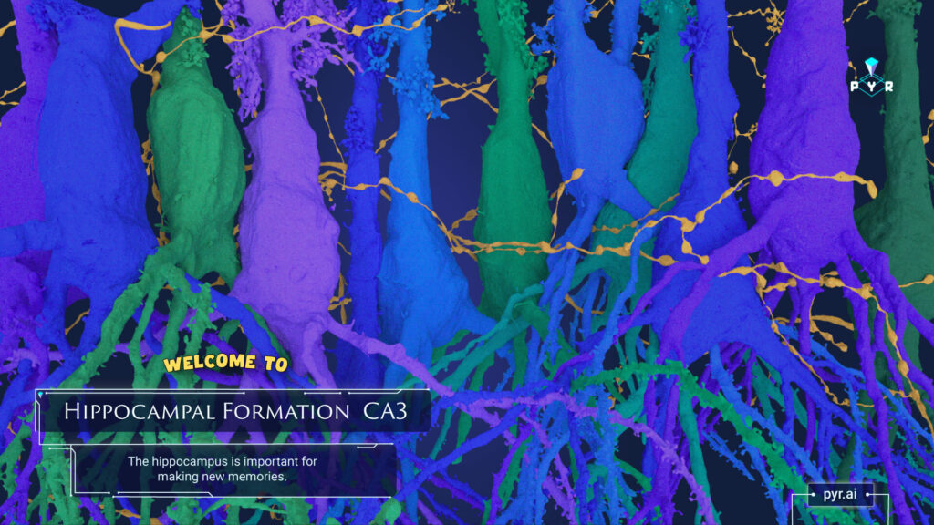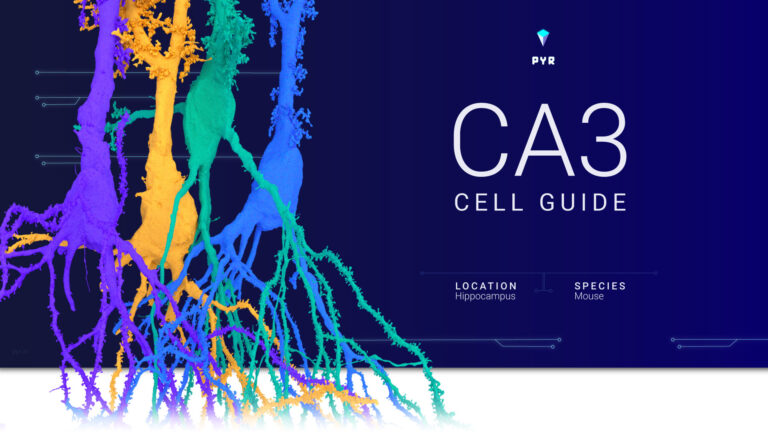
Here is a collection of cell classes found in the CA3 region of mouse hippocampus. To request early dataset access, email support@eyewire.org.
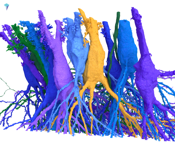
CA3 is a part of the hippocampal formation, a cluster of brain regions involved in learning and memory. It acts in many ways. One is as a pattern-recognition hub, using connections among cells to help form new and process existing memories.
Excitatory Neurons
There are two main excitatory neuron types: Thorny and Athorny.
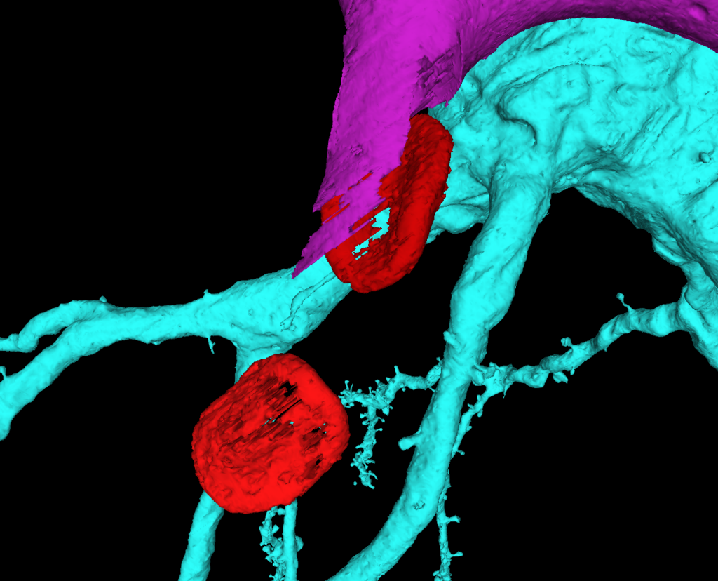
Thorny Pyramidal Neurons due to the “spiny excrescences.” These cells are actively under investigation.
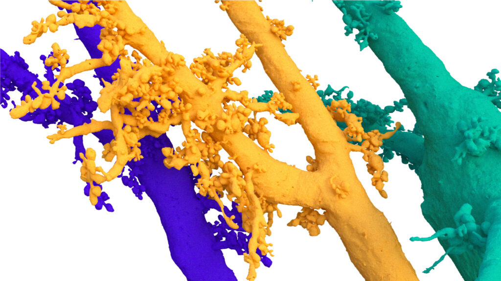
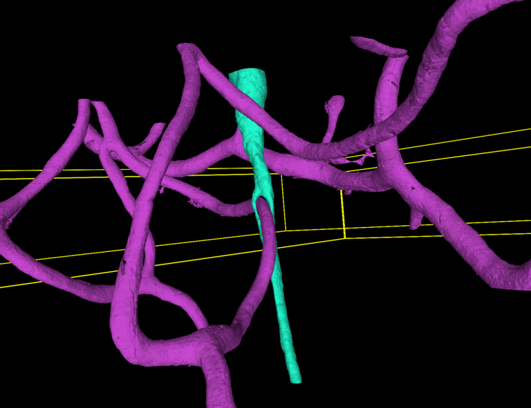
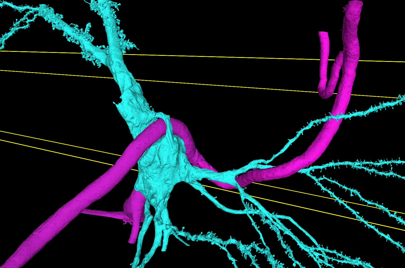
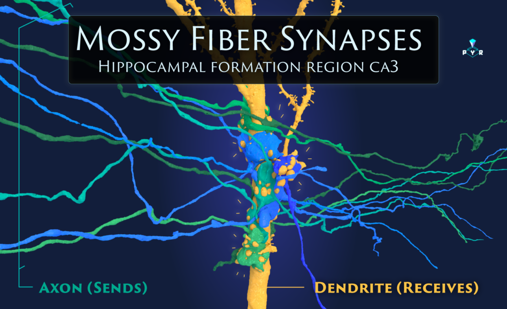
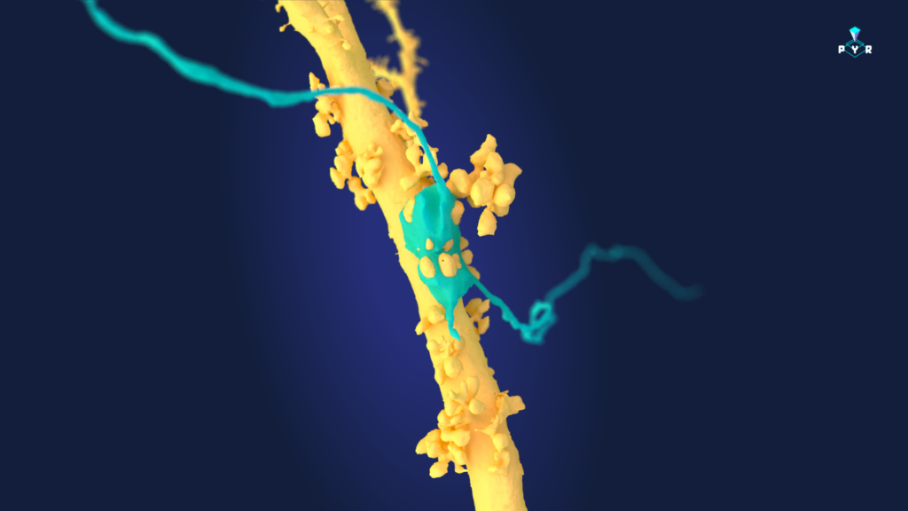
These structures receive input from Mossy Fibers, axons so special that they have their own name. MFs carry info from one part of the brain (the dentate gyrus, also part of the hippocampal formation—aka memory central) to the CA3. The info they bring? It’s crucial for helping your brain learn patterns and make memories.
These axons connect downstream through a unique and giant multi-synaptic structure.
Example of several MF attaching to the throny excrenscences of a Pyr cell: https://spelunker.cave-explorer.org/#!middleauth+https://global.daf-apis.com/nglstate/api/v1/5363463246315520
Their HUGE boutons may attach to several Pyr cells as they travel through CA3. But they will only have one major bouton on an individual Pyr cell (most MF do not synapse more than once with an individual Pyr cell). Mossy fibers (from dentate gyrus granule cells) ONLY form synapses with the thorny CA3 pyramidal cells, specifically at their thorny excrescences.
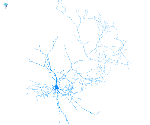
Athorny Pyramidal Neurons.
Excitatory pyramidal neurons. Active investigation.
They receive input from many different sources, including:
- Commissural fibers from the contralateral hippocampus
- Recurrent collaterals from other CA3 neurons
- Schaffer collaterals from CA3 to CA1 neurons
They are named athorny because they lack the spiny excrescences that characterize their counterparts.

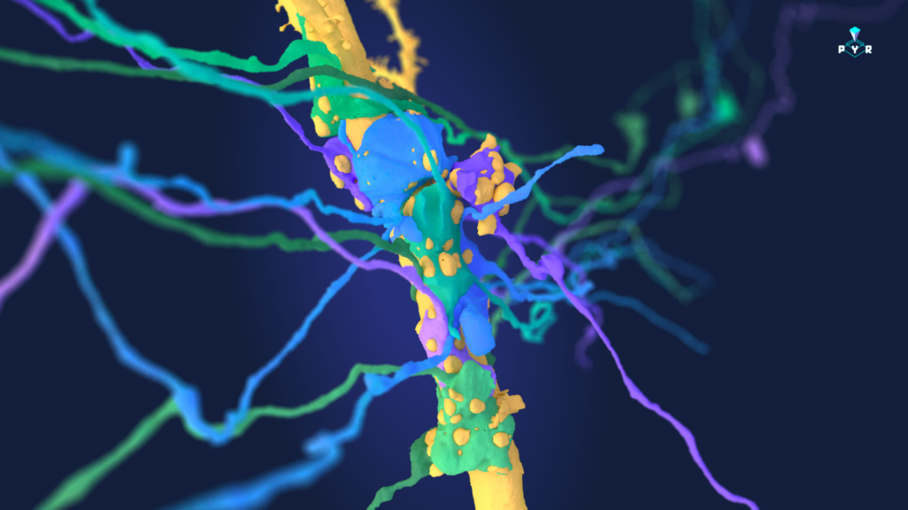
Inhibitory neurons
There are many types of inhibitory neurons in CA3. Find a few below. These cells are under active investigation.


General Oddities
Multiple en passant boutons are converging onto each one of these “inhibitory” spinees.