If you are not familiar with the Spelunker interface, or are new to proofreading, please read the Proofreading 101 with Spelunker blog post before proceeding.
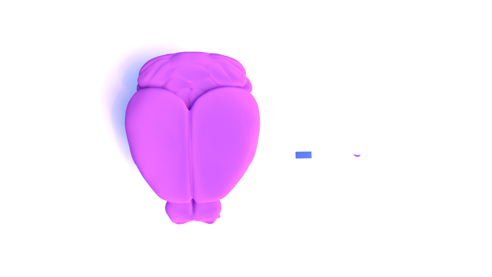
The Minnie dataset is a 1.18mm x 0.8mm x 0.52mm region of cortex within a mouse brain. This is larger than our entire FlyWire dataset! Though it is still spans just a small portion of the mouse brain.
For comparison:
Fly brain: .59 x .34 x .12 mm (source)
Minnie dataset: 1.18mm x 0.8mm x 0.52mm
Mouse brain: 13.2 x 7.4 x 10.4 mm (Allen Institute)
About Pyramidal cells
These excitatory neurons will be our target for proofreading while in Minnie. They are called pyramidal cells due to the pyramid-like shape of their somas (also known as cell bodies).
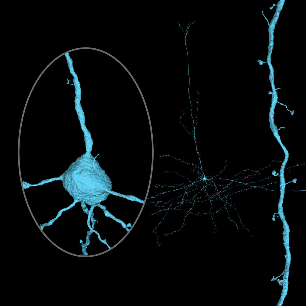
You can identify a pyramidal cell based on it’s morphology, though there are variations between cells, often determined by their location within the dataset.
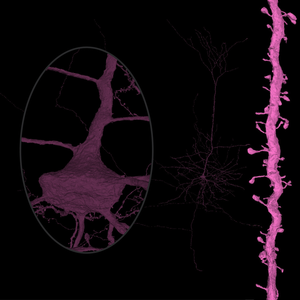
Here is a diagram of the notable parts of a pyramidal cell, though don’t be surprised if cells you are proofreading have some noticeable variation from the cell below.
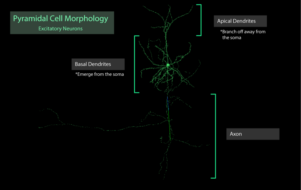
Pyramidal cell parts:
- The soma is in the middle of the cell (looks like a squished ball, or perhaps like a bloated pyramid).
- Basal dendrites are attached to the soma
- Apical dendrites branch off of an apical trunk, which emerges from the apex of the soma
- The axon is much thinner than the dendrites, and reaches in the opposite direction from the apical dendrites
Proofreading Procedure
Because of the large, complex nature of pyramidal cells in Minnie, we have come up with a process for proofreading that ensures a thorough check and minimizes mistakes.
The dendrites and axon have slightly different proofreading procedures, so please make sure you are able to identify the difference between the two.
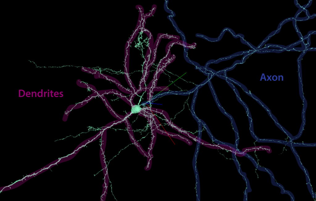
To begin proofreading you will first tackle your cell’s dendritic arbor, and finish by completing the axon.
How to proofread a cell’s dendritic arbor
- Search for mergers and false ends. Check the EM for correction
- Do NOT attach cutoff spine heads
- Extend dendrites through missing sections
- Please spend maximum 10 minutes looking for any one branch extension. If an extension cannot be found within that time you may mark it as “revisit” RV
- Annotations will help you to track your progress. Please use these layer abbreviations and colors when adding annotations:
- soma branch [SB]: branch points where dendrites exit soma, locate 5 microns away from soma, or just soma side of first branch point, whichever is less.
- neuron branch [NB]: branch points for later checking other unvisited branches during proofreading session.
- true end [TE]: ends of a branch
- incomplete end [IE]: branch terminates due to dataset boundaries
- unsure end [UE]: endpoints may stop at this location but the proofreader is not 100% sure
- revisit [RV]: locations where an error was identified or a branch must be extended but could not be fixed in the time frame (10 min) due to bad data, proofreading system timeouts, etc.
- Axon Initial Segment [AIS]: location where the axon branches off (either of soma, or dendrite). Locate 5 microns down axon from soma or branch point.
- other layers that may be pre-populated or added by user:
- centroid [C]: soma centroid
- annotation [annotation]: local layers that are user-added (such as for “breadcrumbs”
**Note: Do not correct small, insignificant mergers that are less than ~5 microns (3x the dendrite diameter)
focus on extending branches instead
Here is an example of a cell with a proofread dendritic arbor, with annotations added
*Note – EM images will not load, please observe the cell in 3D only
How to proofread a cell’s axon
- Search for mergers and remove them
- Search for false ends along the axon in the 3D
- Add segments to each false end until it appears complete
- Please spend maximum 10 minutes looking for any one branch extension. If an extension cannot be found within that time you may mark it as “revisit” RV
- Once all branches are complete, merge added segments to the starter cell
- Annotations:
- axonal initial segment [AIS]: branch point where the axon exits soma
- neuron branch [NB]: branch points for later checking other unvisited branches during proofreading session.
- true end [TE]: end of a branch
- incomplete end [IE]: termination due to dataset boundaries
- unsure end [UE]: endpoint may stop at this location but a proofreader is not 100% sure
- revisit [RV]: location where an error was identified or a branch must be extended but could not be fixed in the time frame (10 min) due to bad data, proofreading system timeouts, etc.
- suspicious [Sus]: Although you see no errors, the branch is making a strange shape, such as a sharp bend in a questionable direction
- difficult area [DA]: Drop an annotation at any point where you had trouble navigating. This creates an easily found place to double check if you notice a branch begins to look merger-like, and is a helpful marker for anyone checking the cell later on
Here is an example of a cell with a proofread axon, with annotations added
*Note – EM images will not load, please observe the cell in 3D only
Here are starter links with annotation layers pre-added:
Sandbox Link (use if you don’t have access to production)
**If you do not see any segmentation in the Sandbox link, you may need to agree to the Terms of Service first. Click here to agree, and then return to the link above.
Proofreading Tips and tricks
- Use the “2” hotkey to turn on and off the 3D meshes frequently. Temporarily turning off the 3D is often useful when looking for extensions in the EM
- You may use “o” to switch between perspective and orthographic views. Test it out to see which you prefer
Tackling Difficulties
Reconstruction errors occur because the AI is having a difficult time finding the correct path through the EM images. Many of these can be difficult for proofreaders to navigate as well.
Let’s go over some typical errors you may run across when proofreading in Minnie.
This slide deck has examples of errors you might find in Minnie:

If you find video examples helpful, we also have an extensive library of help videos for errors found in our FlyWire dataset (pertaining to the brain of Drosophila melanogaster – the fruit fly).
Though the Minnie dataset has differences from the FlyWire dataset, many of the data errors are similar. You can find those videos by clicking below:

What Will My Cell Look Like?
Knowing what your end product should look like can be very helpful when proofreading. While pyramidal cells have the same basic structure, they can also have a lot of variation depending on their location within the cortex.
Here are some resources to help you understand what your cell may look like when completed.
Diagrams from scientific papers
The diagrams below are taken from scientific papers and show a variety of pyramidal cells. You can check out the papers they’re linked to as well for even more information!
1.

In the image above, we will focus on the pyramidal neurons in the left panel. The black portion shows dendrite morphology, while the orange indicates the axon.
Neurons in the upper layers project their axons within the cortex, while neurons in lower layers project their axons through the cortex and into subcortical structures including the striatum, thalamus, brainstem and spinal cord.
*Note: the cells studied in this paper come from a primate, but should have similar features to mouse pyramidal cells.
2.

3.
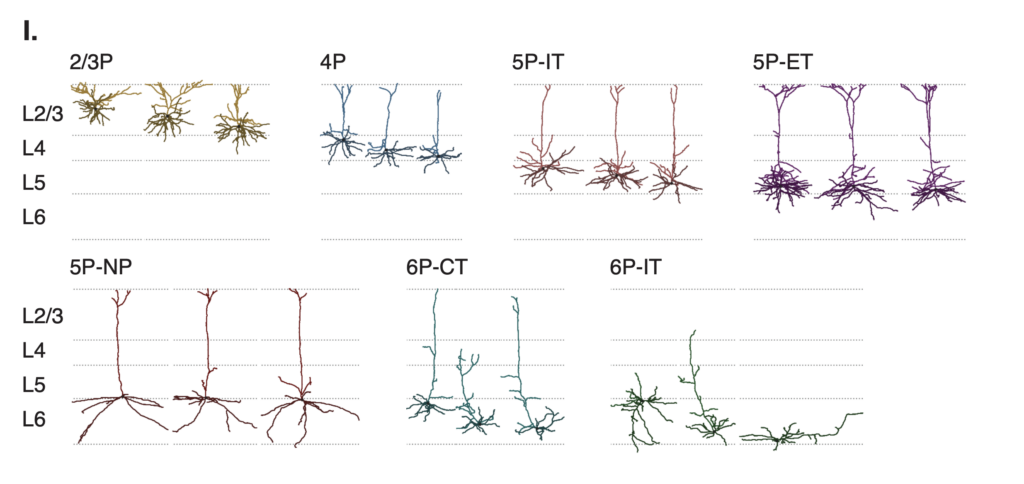
“I. Example morphologies of the expert-defined cell types.”
MICrONS Explorer
The MICrONS Explorer is a scientific tool created to help visualize cells from the Minnie dataset. Use it to explore cell types and familiarize yourself with the mouse cortex.
Here are some good places to get started:
A selection of pyramidal cells from the MICrONS Explorer
A batch of 200 pyramidal cells (caution: some computers may have a hard time with loading)


